Diagnosing
Plaquenil Eye Damage
How Plaquenil Eye Damage Is Diagnosed
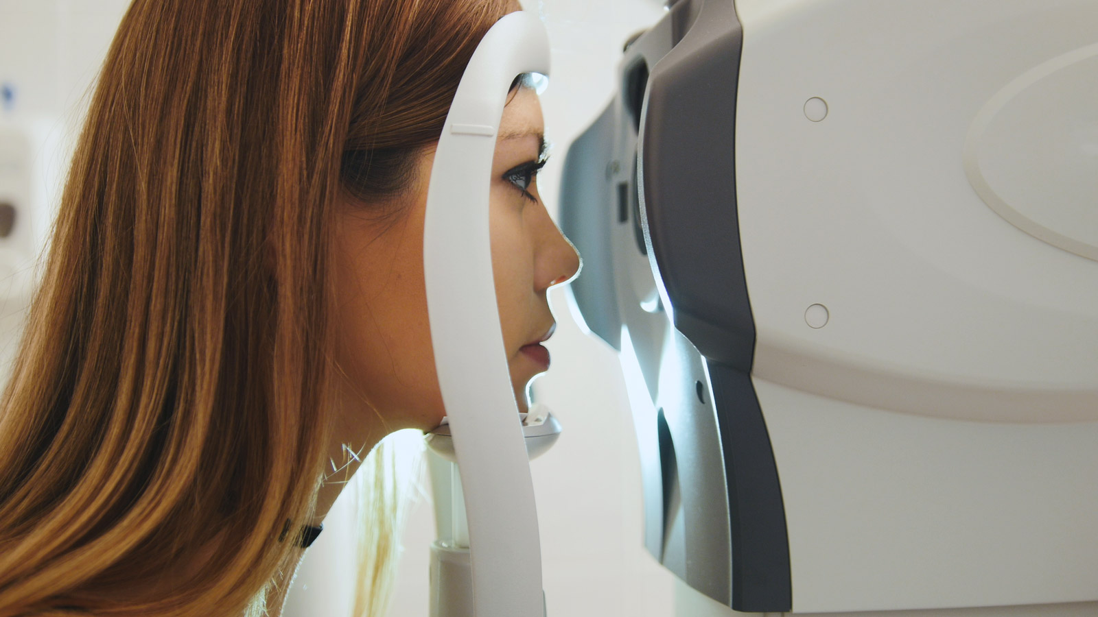
Diagnosing Plaquenil-induced retinal toxicity requires a highly specialized approach that goes far beyond routine vision tests. Because retinal damage from hydroxychloroquine often begins before any visual symptoms appear, detection relies on sensitive, modern imaging and functional testing capable of identifying subtle changes to the retina’s structure and function.
When properly applied, these tests can detect Plaquenil toxicity years before permanent visual loss occurs. However, when physicians or ophthalmologists fail to use the correct screening tools–or delay screening altogether–opportunities for early intervention are missed, and patients may suffer preventable, irreversible vision loss.
Why a Routine Eye Exam Is Not Enough
A standard eye exam typically includes visual acuity testing (the eye chart), intraocular pressure measurement, and ophthalmoscopic evaluation of the retina. While valuable for general eye health, these tools are not capable of detecting early-stage Plaquenil toxicity.
In fact, according to the American Academy of Ophthalmology (AAO), visible signs of damage to the retina–such as bull’s-eye maculopathy–represent an advanced and often untreatable stage of the disease
Similarly, the Amsler grid–a paper test used to check for visual distortions–is not sensitive or specific enough to catch the earliest signs of paracentral vision loss and should not be relied upon as a screening method.

Gold-Standard Tools for Diagnosing Plaquenil Retinopathy
1. Spectral-Domain Optical Coherence Tomography (SD-OCT)
SD-OCT is a non-invasive imaging technology that produces high-resolution, cross-sectional images of the retina, similar to an optical “ultrasound.” It allows for the visualization of individual retinal layers, including the photoreceptors and the retinal pigment epithelium (RPE), where Plaquenil toxicity begins.
- Key Diagnostic Value:
SD-OCT can detect localized thinning or disruption of the outer retinal layers–particularly the ellipsoid zone–before symptoms arise or fundus changes are visible - Adaptation for Asian patients:
Because retinal damage in Asian patients tends to develop in a more peripheral pattern, SD-OCT scans should include wider-angle or pericentral retinal views (e.g., across the vascular arcades) to avoid missing extramacular damage
2. Automated Visual Field Testing (10-2, 24-2, or 30-2)
Automated visual field tests measure light sensitivity across the central and peripheral visual fields, helping to identify areas where vision is diminished–even when the patient is not yet aware of any problems.
- 10-2 Visual Field Test:
This protocol is used in non-Asian patients, targeting the central 10 degrees of vision–where early parafoveal damage usually occurs. It tests 68 points in this region for sensitivity changes. - 24-2 or 30-2 Visual Field Tests:
These protocols test a broader area of vision and are essential for Asian patients, whose retinal damage often appears outside the macula, along the pericentral arcades - Limitation:
Visual field testing is subjective and can vary depending on patient performance, fatigue, or test-retest variability. For this reason, abnormal results should always be confirmed by an objective structural test such as SD-OCT or mfERG.
3. Multifocal Electroretinography (mfERG)
mfERG measures the electrical responses of multiple regions of the retina to visual stimuli, offering a topographic map of retinal function.
- Key Diagnostic Value:
mfERG is capable of detecting functional abnormalities in photoreceptors well before they manifest as visible structural changes. It provides objective evidence of damage and is especially useful for confirming ambiguous visual field findings - Limitations:
The test requires highly trained personnel and specialized equipment, which is generally available only at larger ophthalmology clinics or academic centers.
4. Fundus Autofluorescence (FAF)
FAF imaging captures natural fluorescence from the retina, allowing clinicians to visualize the metabolic state of the retinal pigment epithelium (RPE).
- Early Detection:
Areas of increased autofluorescence may signal early RPE stress or impending damage, while dark areas reflect RPE cell death. These changes can precede thinning on OCT or abnormalities on visual field testing - Topographic Mapping:
FAF provides a wide-field view of the retina, making it especially helpful in patients with atypical damage patterns, including those with Asian heritage.
Losing your vision to Plaquenil is a devastating and irreversible outcome.
If you or a loved one has suffered eye damage from this medication, contact us today for a free and confidential case evaluation to understand your legal options.

